(Peer-Reviewed) Ferritin as a diagnostic, differential diagnostic, and prognostic marker for immune-related adverse events
Weihong Zhang 张维红 ¹, Yuan Meng ¹, Lin Yang 杨琳 ², Meng Shen 沈梦 ¹, Li Zhou 周莉 ¹, Runmei Li 李润美 ¹, Yang Wang 王扬 ¹, Weijiao Du 杜伟娇 ¹, Yanjuan Xiong 熊艳娟 ¹, Ying Han 韩颖 ¹, Xinwei Zhang 张新伟 ¹, Liang Liu 刘亮 ¹, Xiubao Ren 任秀宝 ¹
¹ Department of Immunology and Biotherapy, Key Laboratory of Cancer Immunology and Biotherapy, Tianjin Medical University Cancer Institute and Hospital, National Clinical Research Center for Cancer, Key Laboratory of Cancer Prevention and Therapy, Tianjin, Tianjin's Clinical Research Center for Cancer, Tianjin 300060, China
中国 天津 天津市肿瘤免疫与生物治疗重点实验室 天津医科大学肿瘤医院肿瘤研究所 国家肿瘤临床研究中心 天津市肿瘤防治重点实验室 国家肿瘤临床医学研究中心
² State Key Laboratory of Experimental Hematology, Peking Union Medical College, Institute of Hematology and Blood Diseases Hospital, Chinese Academy of Medical Sciences, Tianjin 300020, China
中国 天津 北京协和医学院 实验血液学国家重点实验室 中国医学科学院血液学研究所
Objective
Distinguishing immune-related adverse events (irAEs) caused by immune checkpoint inhibitors (ICIs) from the AEs caused by chemotherapy, targeted therapy, or infection is highly difficult. This study offers new insights into evaluating the diagnosis, differential diagnostic, and prognostic value of ferritin for irAEs induced by ICIs.
Methods
From December 1, 2018, to April 1, 2019, we examined 318 patients with malignant tumors who received serum ferritin monitoring. The cohort comprised 231 patients treated with PD-1 inhibitor or combination with chemotherapy, and 87 patients treated with chemotherapy. Of the 231 patients, 90 had irAEs (irAE group), 70 had non-irAEs (non-irAE group), 67 had no AEs (no irAE-non irAE group), and 4 had unclassified AEs. In the 87 patients, 60 had AEs (AE group), and 27 had no AEs (no AE group). Statistical analyses were conducted with nonparametric Mann-Whitney tests.
Results
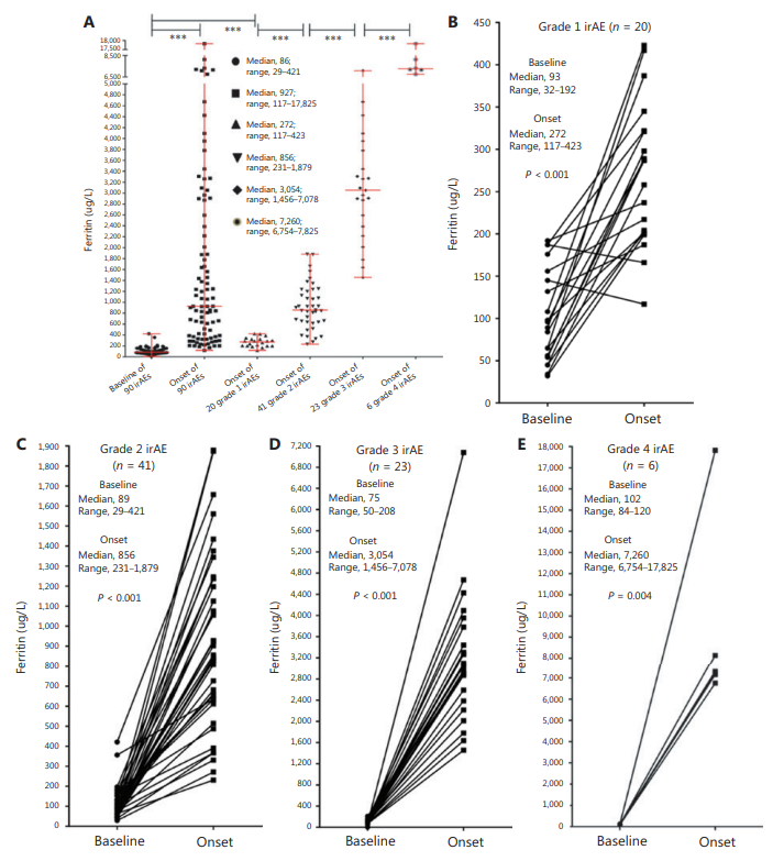
At the onset of AEs in the irAE group, ferritin (normal range, 35–150 µg/L) rose to a median of 927 µg/L (range, 117–17,825 µg/L) from 86 µg/L at baseline (range, 29–421 µg/L) (P < 0.001). Ferritin levels at the onset of AEs in the irAE group were significantly higher than those in the non-irAE group (median, 81 µg/L; range, 32–478 µg/L) (P < 0.001) and the AE group (median,
103 µg/L; range, 23–712 µg/L) (P < 0.001). After treatment in the irAE group, ferritin ntinuously decreased to a normal range in recovered patients, showed no significant changes in stable patients, and continued to rise in patients who died.
Conclusions
Ferritin can be used as a diagnostic, differential diagnostic, and prognostic marker for irAEs in patients treated with ICIs.
High-resolution tumor marker detection based on microwave photonics demodulated dual wavelength fiber laser sensor
Jie Hu, Weihao Lin, Liyang Shao, Chenlong Xue, Fang Zhao, Dongrui Xiao, Yang Ran, Yue Meng, Panpan He, Zhiguang Yu, Jinna Chen, Perry Ping Shum
Opto-Electronic Advances
2024-12-16
Ultra-high-Q photonic crystal nanobeam cavity for etchless lithium niobate on insulator (LNOI) platform
Zhi Jiang, Cizhe Fang, Xu Ran, Yu Gao, Ruiqing Wang, Jianguo Wang, Danyang Yao, Xuetao Gan, Yan Liu, Yue Hao, Genquan Han
Opto-Electronic Advances
2024-10-31


